Rushil Srirambhatla
Biomedical AI · Computer Vision · Medical Imaging

Rushil Srirambhatla

I develop machine learning methods for medical imaging, from multimodal 3D CT generation to robust neuroimaging analysis and disease-related representation learning. I am completing a B.S. in Applied Mathematics & Statistics and Cognitive Science at Johns Hopkins University (December 2026).
Computer Vision3D generative modelingMedical ImagingMultimodal clinical AI
Rushil Srirambhatla

Research at a Glance

Generative Medical AI

Text-conditioned 3D CT synthesis and clinically guided image generation.

Medical Computer Vision

Robust brain extraction, image segmentation, and quantitative 3D analysis.

Neuroimaging & Representation Learning

Brain-age prediction, MRI reconstruction, and imaging biomarkers of disease.

Reliable Biomedical ML

Reproducible benchmarks, interpretable models, and clinically meaningful evaluation.

News

Recent research updates, acceptances, and conference announcements.

CT brain-extraction benchmark pending acceptance

Our multi-site evaluation of seven automated CT brain-extraction methods across 5,062 scans is pending acceptance at BMC Medical Imaging.

Stroke 2027 oral abstract accepted

Co-authored work on insurance coverage gaps among working-age U.S. stroke survivors was accepted for an oral presentation at the 2027 International Stroke Conference.

3D MRI autoencoder accepted at ECCV workshop

Our paper on memory-efficient, skip-connection-free whole-brain MRI reconstruction was accepted to the ECCV 2026 AI4M3D Workshop.

Research & Publications

Selected work in medical computer vision, 3D imaging, multimodal learning, and computational genomics.
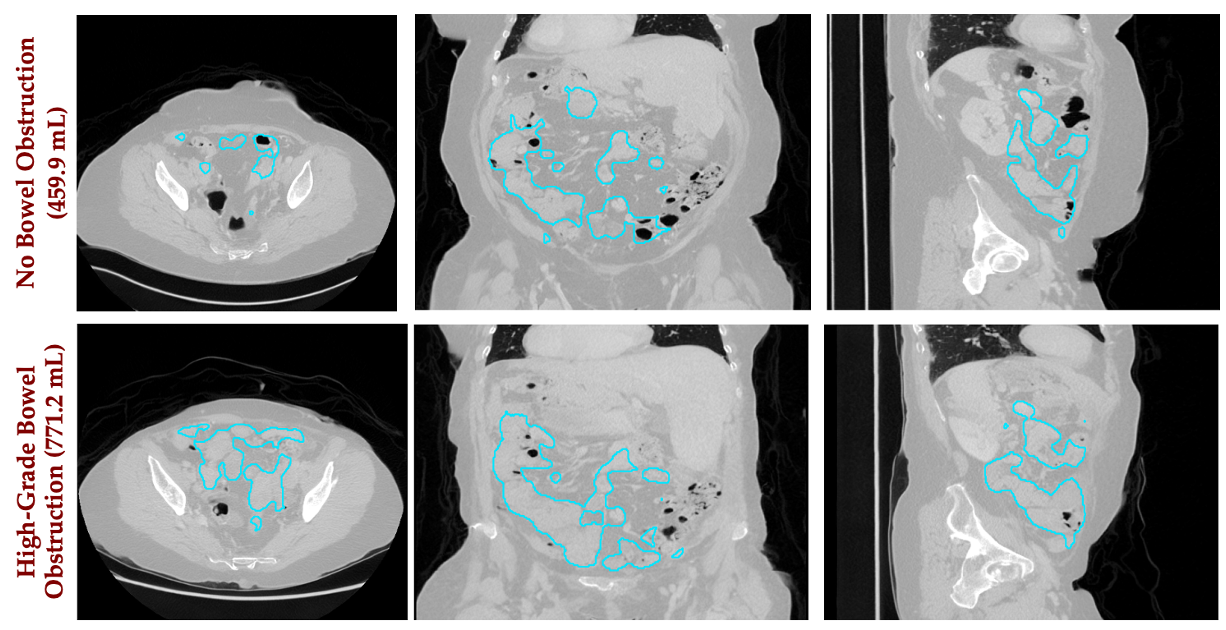
Representative clinical-report-conditioned 3D CT synthesis result

Clinical-report-conditioned 3D CT synthesis

Rushil Srirambhatla, Akshay Chaudhari, David Larson · Stanford AIDE Lab

Fine-tuned three MAISI diffusion U-Nets with ClinicalT5 cross-attention, comparing impression-only, full-report, and MedGemma-structured reports on matched study splits. Built preprocessing for 25,489 Merlin CT studies.

Key finding: Structured conditioning improved BiomedCLIP text–image alignment by ~42%; fixed-noise counterfactual experiments revealed limitations in anatomical control.
CodePresentation PDF
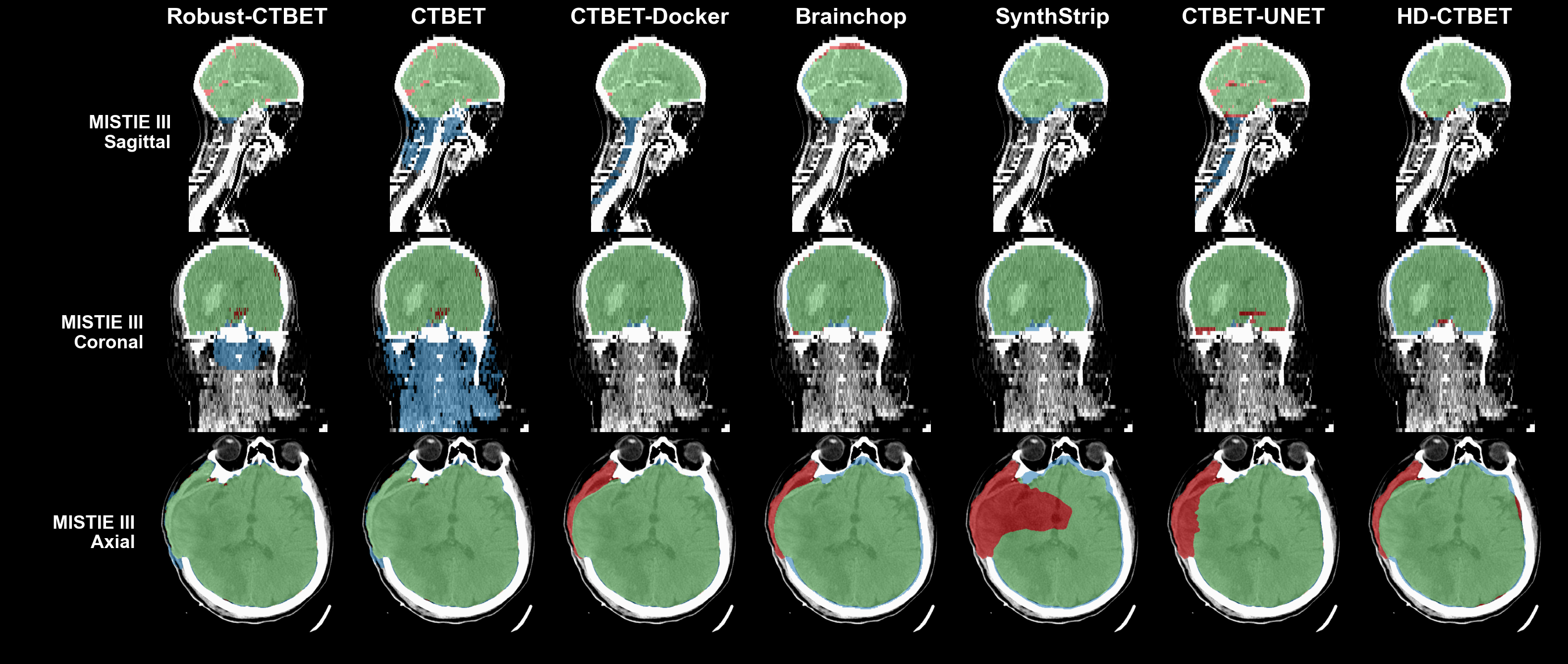
Comparison of seven CT brain-extraction methods across sagittal, coronal, and axial views

Benchmarking Automated CT Brain Extraction Methods Across 5,000 Scans

Rushil Srirambhatla, Nathan Walborn, Daniel Hanley, John Muschelli

Developed Robust-CTBET and benchmarked seven brain-extraction methods on 5,062 CT scans from 499 patients at 78 sites, assessing registration, volumetrics, and downstream utility.

Key finding: Robust-CTBET outperformed six competing methods, achieving Dice ≈ 0.98 and a 0.30% scan-level failure rate.
Code & resultsAbstract
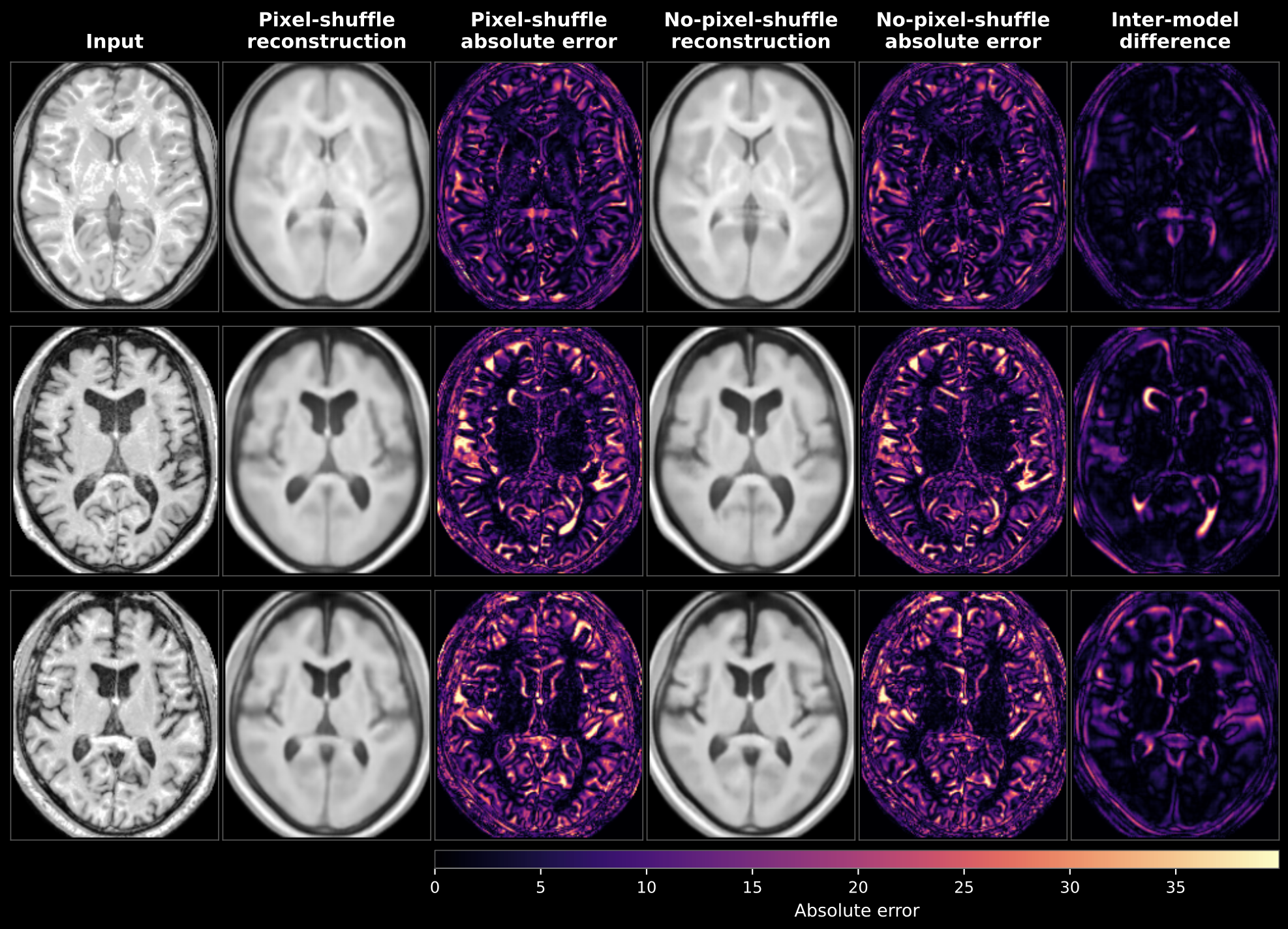
Figure for Memory-Efficient Skip-Connection-Free 3D Autoencoder for Whole-Brain MRI Reconstruction

Memory-Efficient Skip-Connection-Free 3D Autoencoder for Whole-Brain MRI Reconstruction

Rushil Srirambhatla, Helmet T. Karim, Jacques-Yves Campion

Designed a skip-connection-free 3D autoencoder with a 512-dimensional bottleneck and pixel-shuffle decoder for full-volume T1-weighted MRI reconstruction, evaluated across 40,841 scans.

Key finding: 19.6% less peak GPU memory, 10.4% faster forward/backward execution, and 13.4% fewer parameters versus the no-pixel-shuffle baseline.
Code & resultsPaper
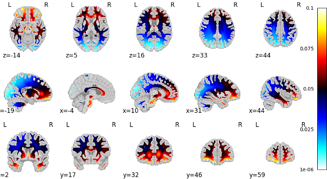
Brain maps showing disease-related brain-age effects across axial, sagittal, and coronal views

Deep Learning-Derived Brain Age from 40,000+ MRI Scans Reveals Disease-Specific Aging Trajectories

Jacques-Yves Campion* and Rushil Srirambhatla*, Carmen Andreescu, et al. · *Equal contribution

Developed brain-age prediction pipelines using harmonized 3D MRI and studied disease-associated deviations in predicted brain age across multi-cohort clinical imaging data.

Key finding: 3.14-year held-out test MAE in the brain-age prediction study.
Code & results
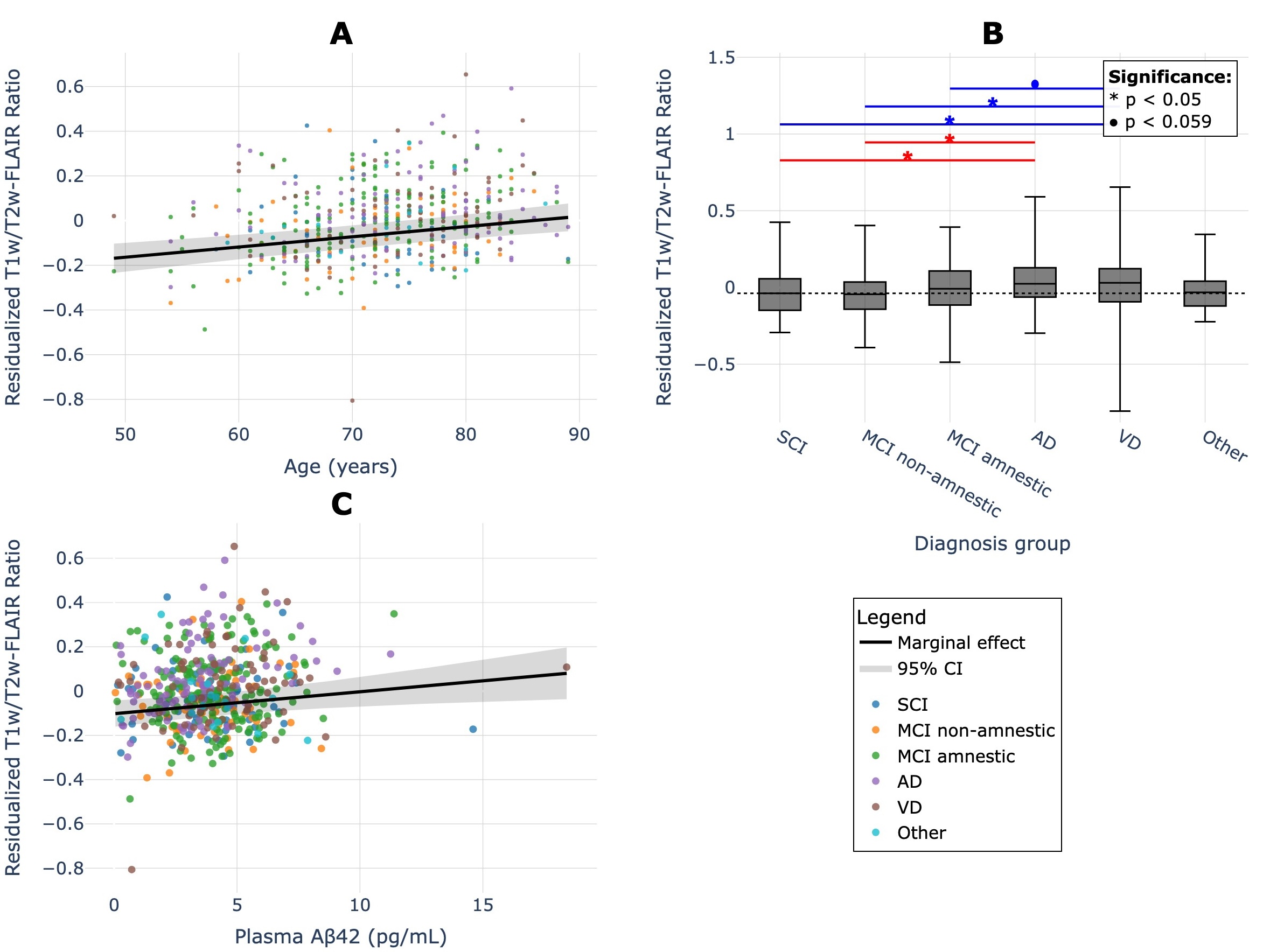
White matter hyperintensity T1w/T2w-FLAIR ratio analysis figure

Altered T1w/T2w-FLAIR Ratio in White Matter Hyperintensities as an Indicator of Structural Integrity Loss

Rushil Srirambhatla, Jacques-Yves Campion, Thomas Desmidt, et al.

Integrated LST-AI lesion segmentation, ANTs preprocessing, and relative T1w/T2w-FLAIR measures in 576 participants to investigate Alzheimer’s disease, vascular dementia, and amyloid biomarkers.

Key finding: White matter hyperintensity (WMH) T1w/T2w-FLAIR ratios were significantly higher in individuals with Alzheimer’s disease and vascular dementia compared with those with subjective cognitive impairment, suggesting disease-associated alterations in white matter tissue integrity.
Code & resultsPreprint
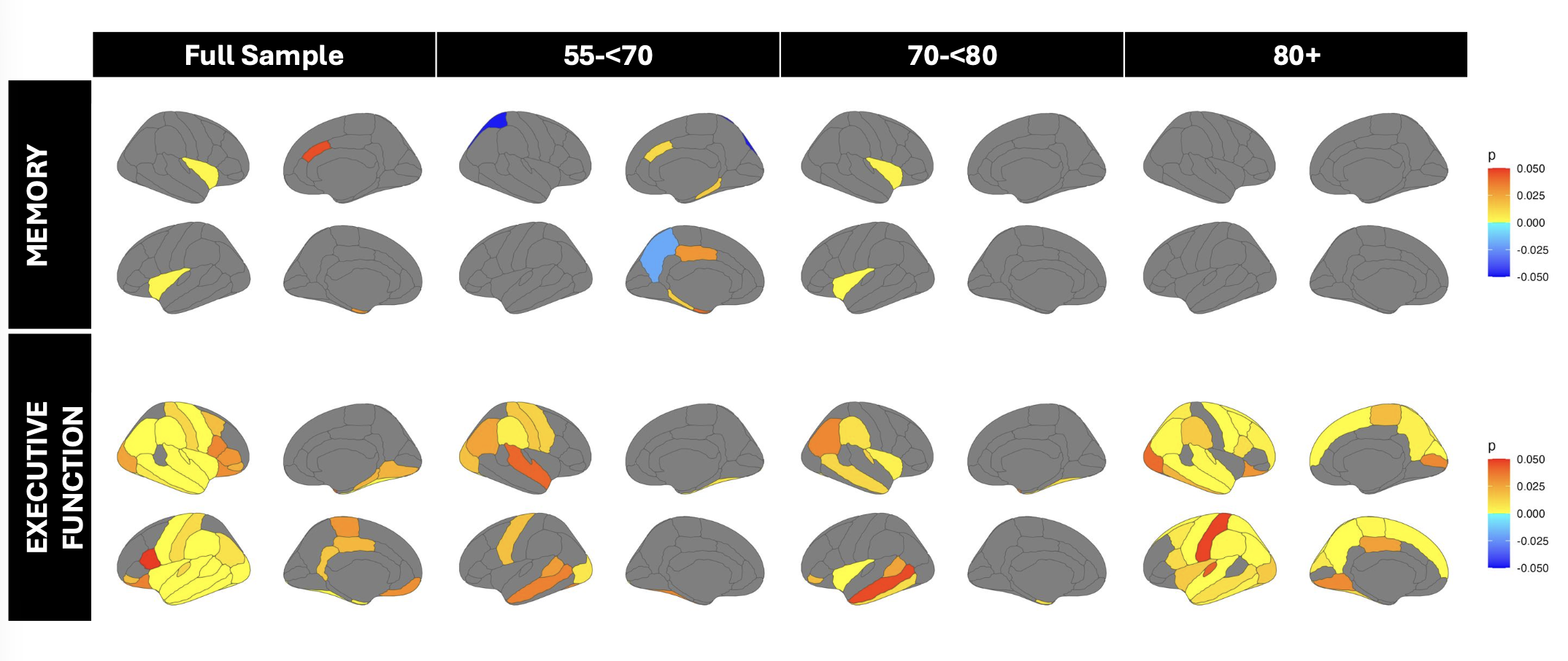
Cortical surface maps comparing cognitive performance across age groups in the ADNI dataset

Cortical Thickness Signatures of High vs. Low Cognitive Performers in the ADNI Dataset

Rushil Srirambhatla, Yue Hong, Yiwen Zhang, David H. Salat

Analyzed FreeSurfer-derived cortical thickness across 68 regions in relation to cognition using age-stratified regression, ANCOVA, and FDR correction.

Key finding: Executive-function associations were most extensive among adults aged 80 and older.
Code & results
Illustration accompanying The Black Box Dilemma commentary on human–AI collaboration

The Black Box Dilemma: Challenges in Human-AI Collaboration in ML-CDSS

Rishab Jain, Rushil Srirambhatla, John Kessler, Ram Goel

A commentary on interpretability and human–AI collaboration in machine-learning-based clinical decision support.
Published commentary
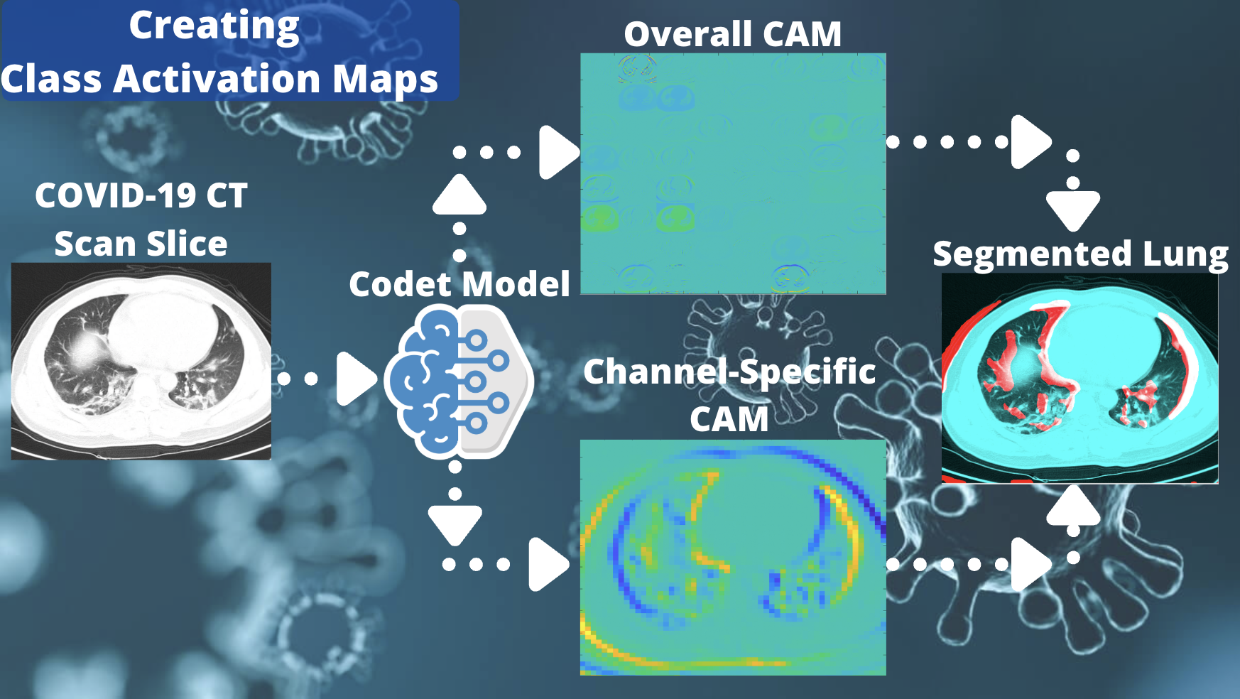
COVID-19 CT classification and model-interpretability figure

Prediction of COVID-19 Diagnosis from Healthy and Pneumonia CT Scans Using Convolutional Neural Networks

Rushil Srirambhatla, Helmet T. Karim

Developed an interpretable CNN-based approach to classifying COVID-19 from CT imaging, incorporating lung-focused preprocessing and activation-map visualization.

CodePreprint

Selected presentations

A selection of first-author presentations and collaborative conference contributions. Click a thumbnail when a public abstract or presentation page is available.
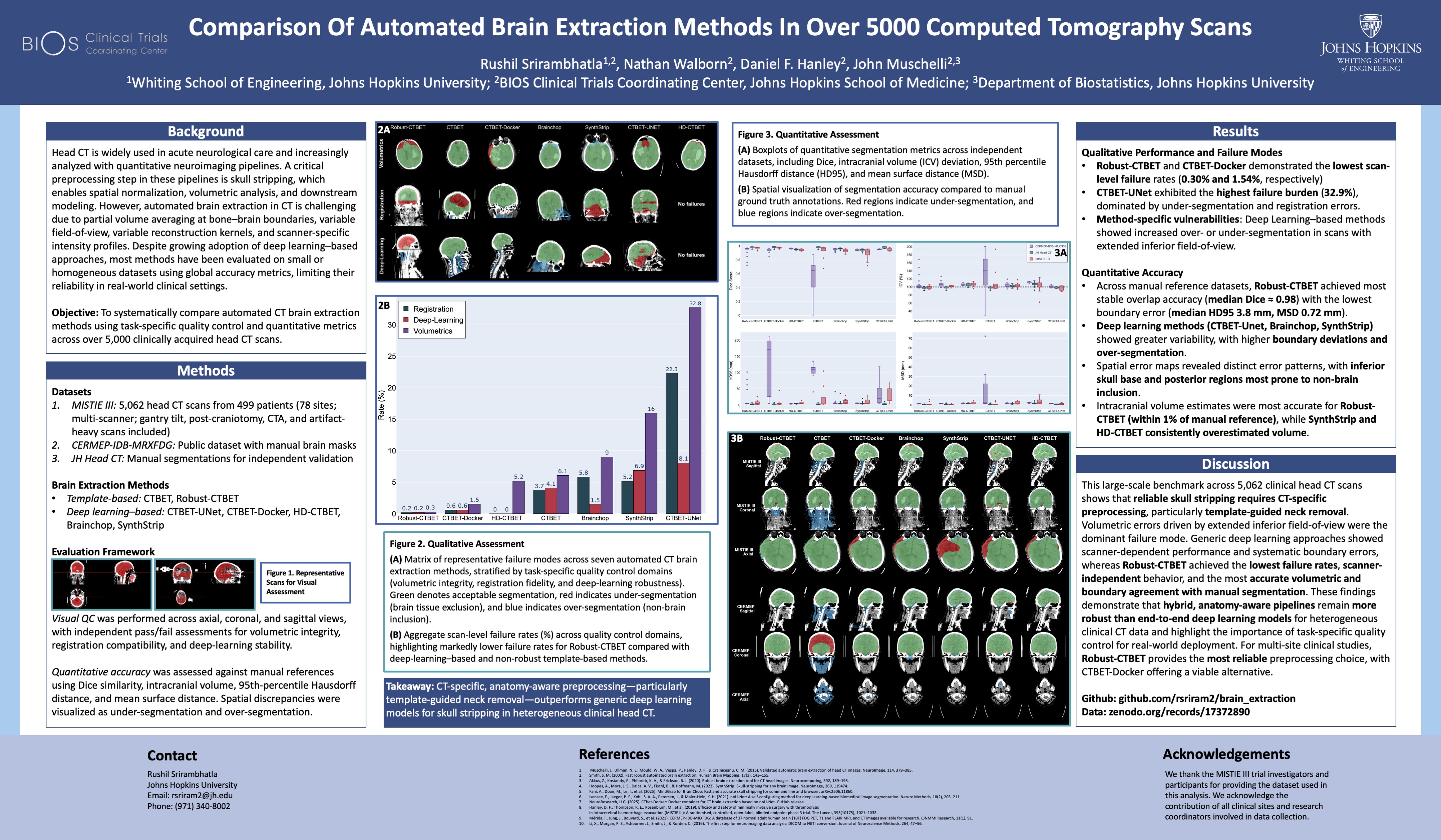
Presentation thumbnail for Comparison of Automated Brain Extraction Methods in Over 5,000 Computed Tomography Scans
2026 · International Stroke Conference

Comparison of Automated Brain Extraction Methods in Over 5,000 Computed Tomography Scans

First author · Conference abstract

Abstract DOI ↗
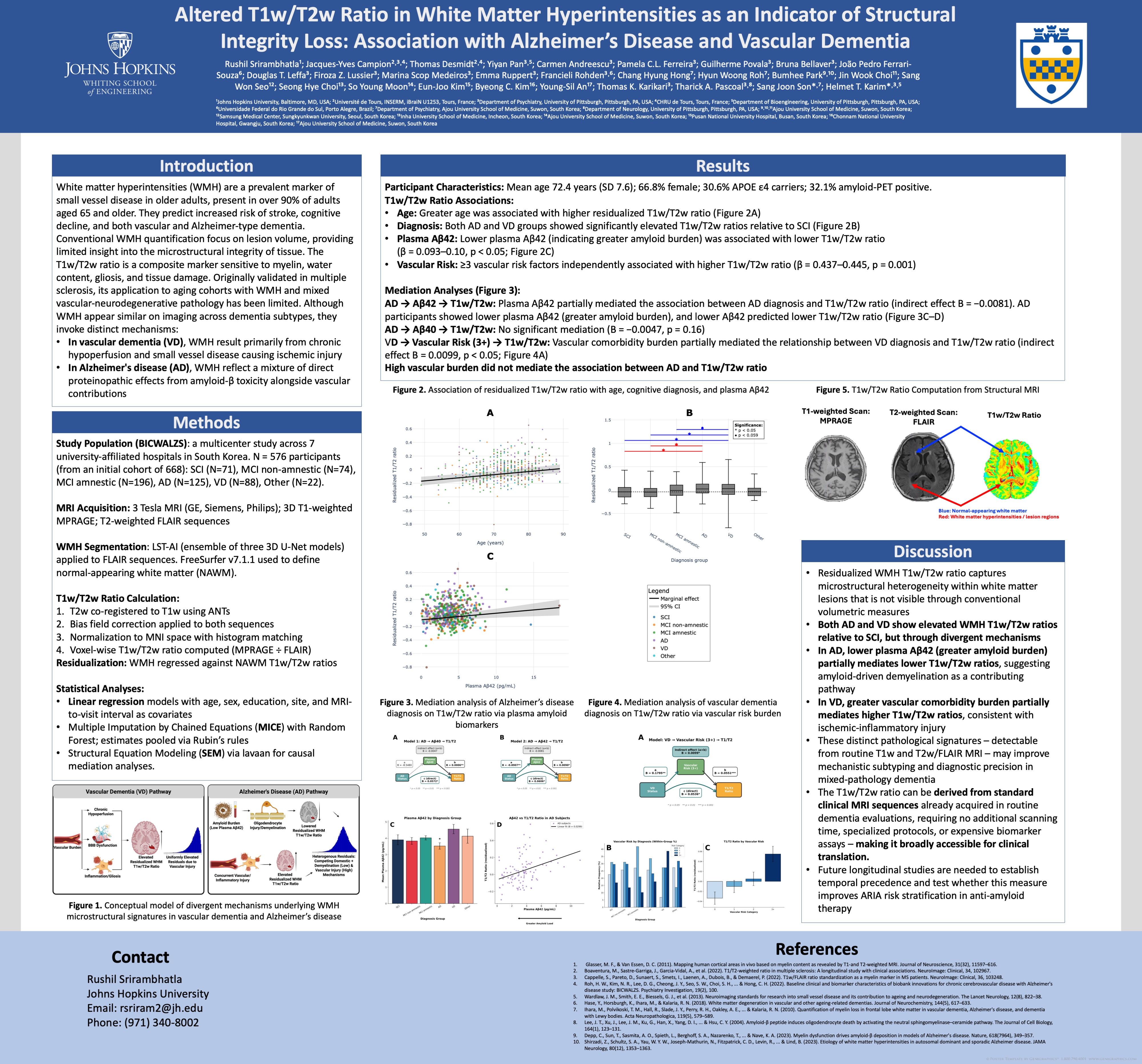
Presentation thumbnail for Altered T1w/T2w Ratio in White Matter Hyperintensities as an Indicator of Structural Integrity Loss
2026 · AAIC / Society of Biological Psychiatry (SOBP)

Altered T1w/T2w Ratio in White Matter Hyperintensities as an Indicator of Structural Integrity Loss

First author · Poster presentation

AAIC Poster ↗
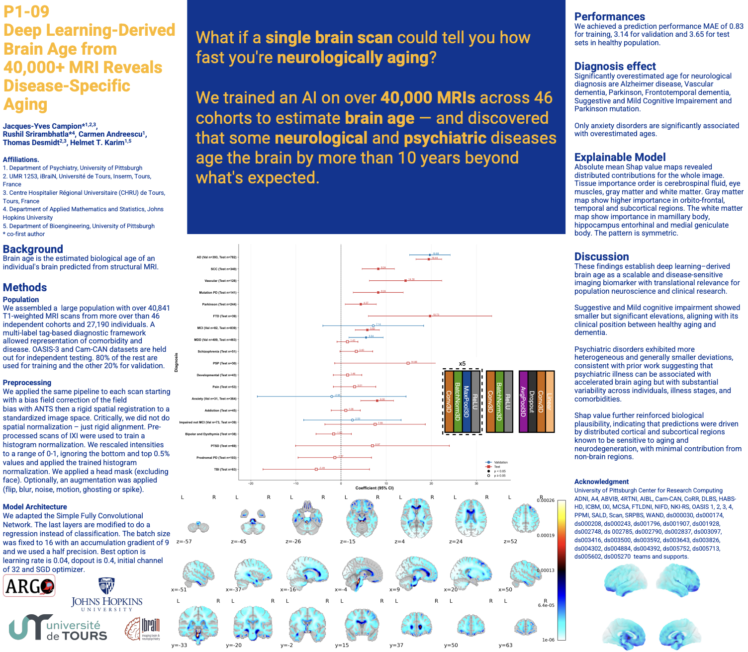
Presentation figure for Deep Learning–Derived Brain Age from 40,000+ MRI Scans Reveals Disease-Specific Aging Trajectories
2026 · AAIC / Society of Biological Psychiatry (SOBP)

Deep Learning–Derived Brain Age from 40,000+ MRI Scans Reveals Disease-Specific Aging Trajectories

Co-first author · Poster presentation

AAIC Poster ↗
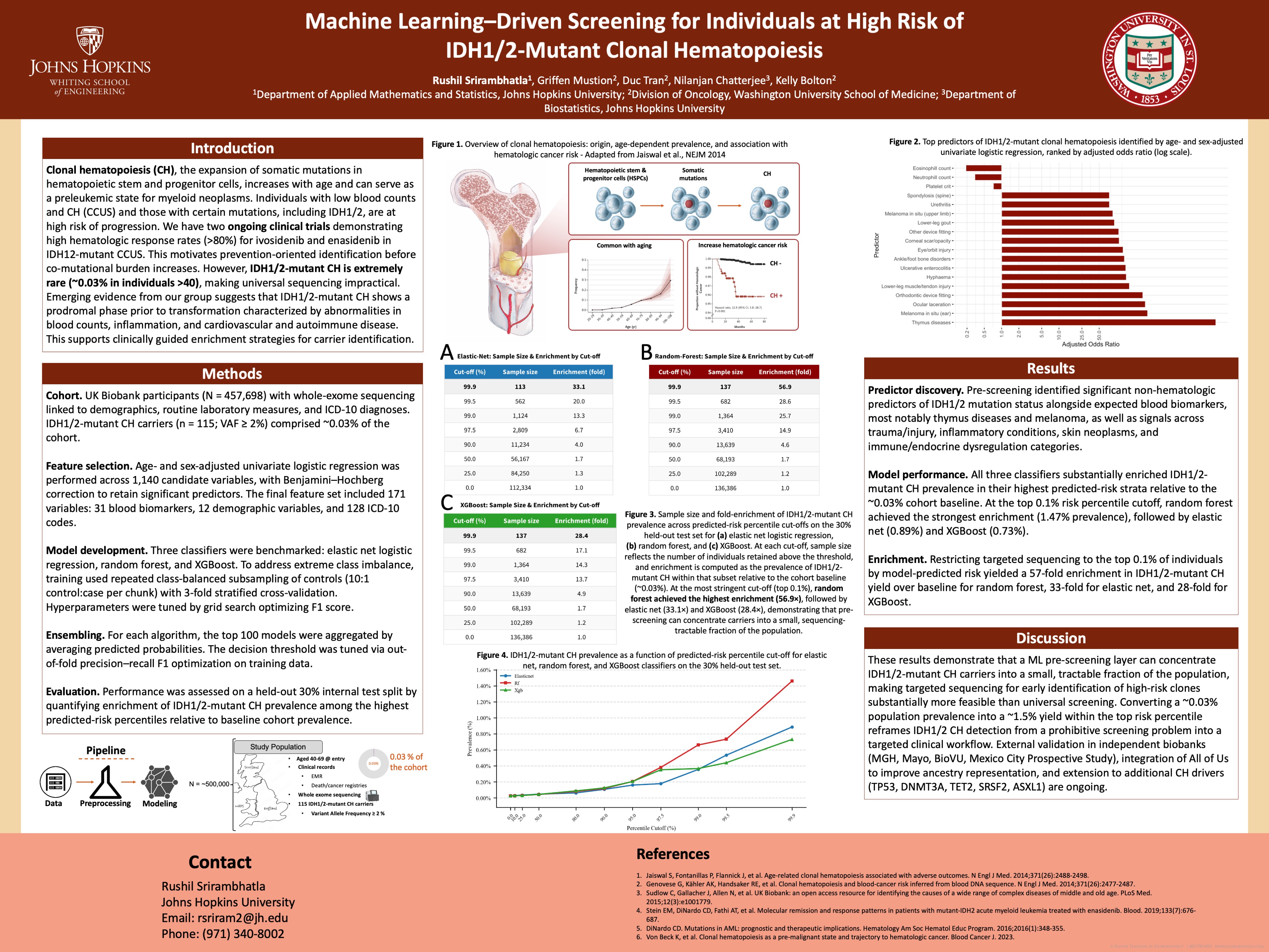
Presentation thumbnail for Machine Learning–Driven Screening for Individuals at High Risk of IDH1/2-Mutant Clonal Hematopoiesis
2026 · 15th International Myeloid Workshop

Machine Learning–Driven Screening for Individuals at High Risk of IDH1/2-Mutant Clonal Hematopoiesis

First author · Poster presentation

Poster PDF ↗
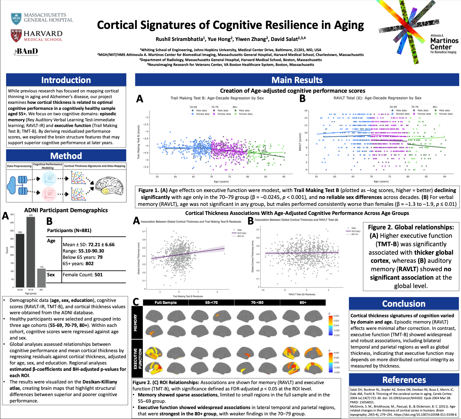
Presentation thumbnail for Cortical Thickness Signatures of High vs. Low Cognitive Performers in the ADNI Dataset
2025 · Mass General Brigham Neuroscience Day

Cortical Thickness Signatures of High vs. Low Cognitive Performers in the ADNI Dataset

First author · Poster presentation

Poster PDF ↗
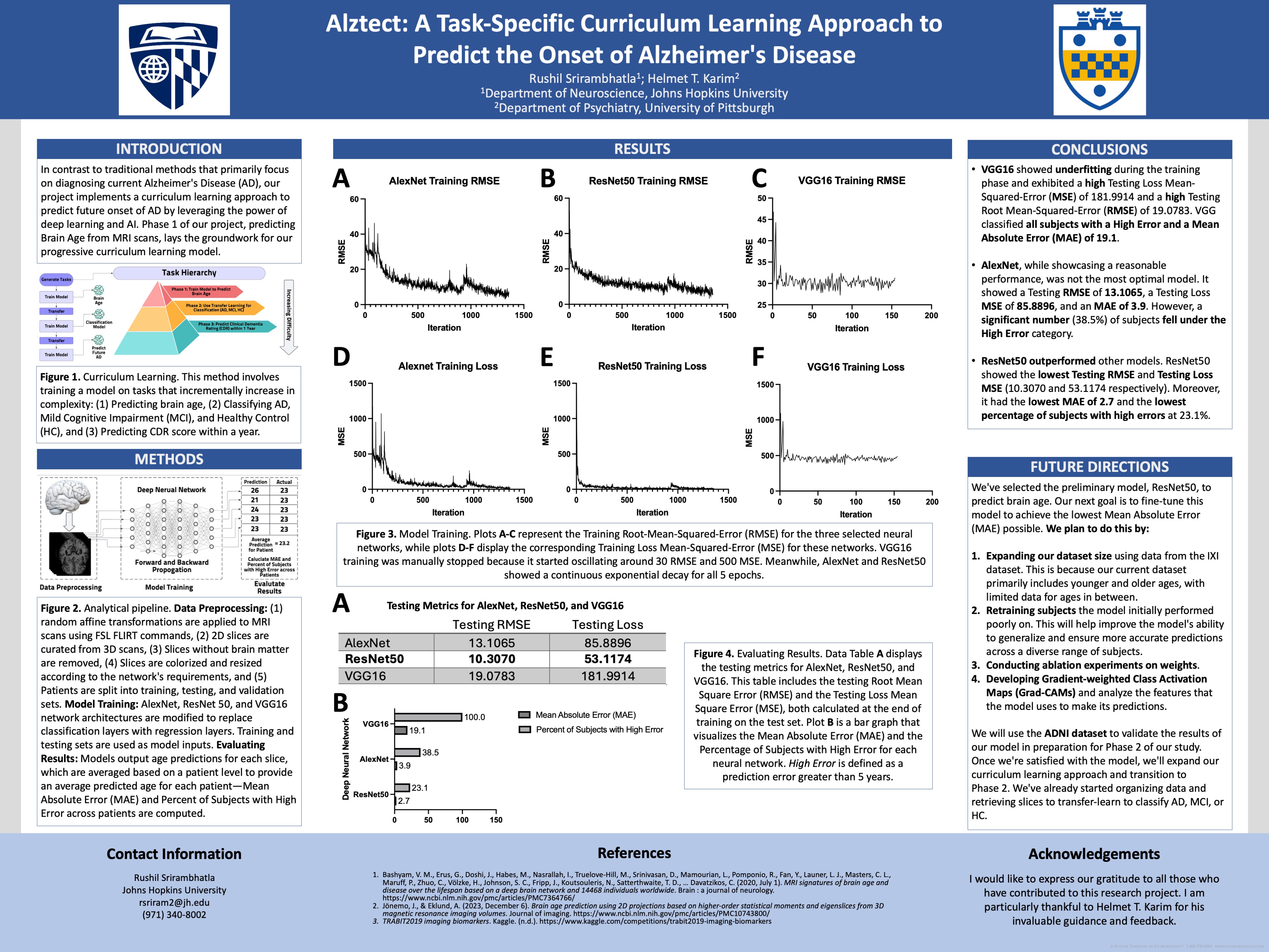
Presentation thumbnail for A Task-Specific Curriculum Learning Approach to Predict the Onset of Alzheimer's Disease
2024 · Pitt Psychiatry Research Day / Johns Hopkins DREAMS

A Task-Specific Curriculum Learning Approach to Predict the Onset of Alzheimer's Disease

First author · Poster presentation

Poster ↗
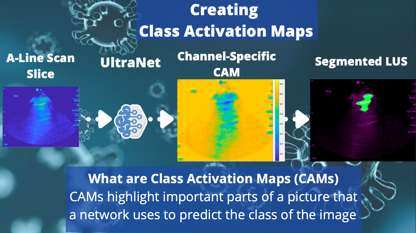
Presentation thumbnail for UltraNet lung ultrasound A-line and B-line differentiation
2023 · Point-of-Care Ultrasound Research Seminar

UltraNet: Differentiating A-Line and B-Line Lung Ultrasound Using Deep Learning

First author · Research seminar presentation

2027 · International Stroke Conference · Accepted oral abstract

Persistent Insurance Coverage Gaps Among Working-Age United States Stroke Survivors: No Improvement From 1999 Through 2023

Co-author · Oral abstract accepted

Research experience

Research appointments across medical image analysis, generative modeling, neuroimaging, and computational genomics.

Machine Learning Research Intern

Stanford University

AIDE Lab
2026–PresentMentors: Akshay Chaudhari, David Larson
3D CT Generation · Multimodal AI
Machine Learning Researcher

Johns Hopkins University

BIOS Clinical Trials Coordinating Center
2024–PresentMentor: John Muschelli
CT Brain Extraction · Benchmarking
Deep Learning Researcher

UPMC

ARGO Group
2021–PresentMentor: Helmet T. Karim
Brain Aging · MRI Representation Learning
Neuroimaging Researcher

Massachusetts General Hospital

BAnD Lab
2025–PresentMentor: David H. Salat
Cortical Morphometry · Cognition
Computational Cancer Genomics Researcher

WashU School of Medicine

Bolton Lab
2025Mentor: Kelly Bolton
Rare-Variant Screening · Predictive ML
Clinical Research Data Specialist

University of Miami

Miller School of Medicine
2023–2025Mentors: John Muschelli, Trishul Siddharthan, Paul Blair
Lung Ultrasound · Computer Vision

Contact

For research discussions, collaborations, or questions about my work: